
.png)
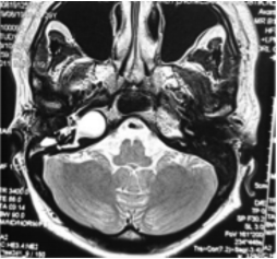
Problem/objective: Multiple differential pathologies can occur at the petrous apex. Clinicians must be highly suspicious of temporal bone pathology to diagnose lower motor neuron facial nerve palsy, particularly in patients with hearing impairments. The clinical presentation of a petrous apex lesion is variable, depending on the involvement of intimately adjacent structures. It cannot be diagnosed accurately from clinical findings alone. Hence, computed tomography and magnetic resonance imaging provide complementary data for more definitive characterization. Here, we described a case of massive petrous apex cholesteatoma in a young adult female that had been misdiagnosed with Bell’s palsy for years. We aimed to highlight a safe treatment approach that could preserve hearing.
Methodology: We used a supralabyrinthine approach for an endoscopic-assisted transmastoid removal of the petrous apex lesion. Results: Post-operative residual hearing was preserved and healing was good.
Conclusion: Endoscopy approach could be a less invasive and effective way in treating PBC without compromising the ultimate aim of complete cholesteatoma removal.